Login
 USD $
USD $
 AUD AUD
 CAD CAD
 GBP £
GBP £ EUR €
EUR €
All Categories
(0) My Cart (0)
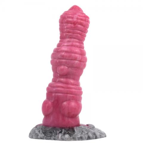
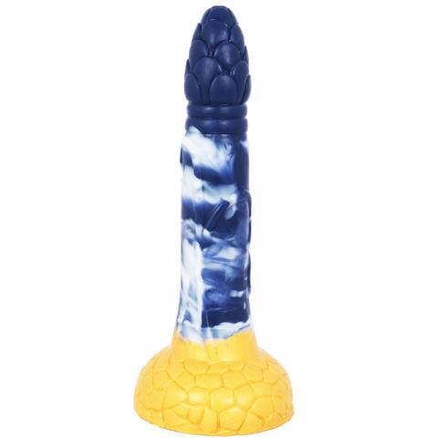
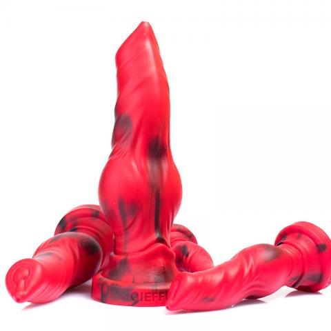
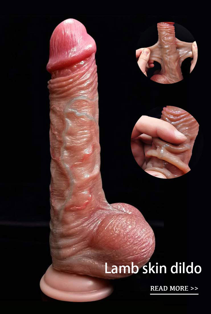
Lambskin Dildo
New Arrival
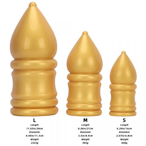
Mushroom Dildo
Cheap
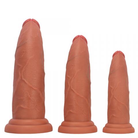
Huge Dildo
Penis Pumps
Masturbation Cup
Chastity Cage
Pocket Pussy
pleated filter cartridge factory
high flow filter cartridge
large flow filter cartridge
membrane pleated filter cartridge
capsule filter suppliers
capsule filter 0.2 micron
capsule filter price
capsule filter
water filter cartridge


The most detailed 3D model of HIV comes out in history.
3D picture of HIV
By far the most detailed 3D model of HIV has won the first prize in the graphic category of the 2010 International Science and Engineering Visualization Challenge. A Russian research team led by Ivan Konstantinov analyzed data from more than 100 scientific journals and then used digital means to present HIV as realistically as possible. This diagram is made in two colors, orange and gray, representing HIV and an immune cell, respectively. The picture shows a terrible picture of the virus attacking and invading the cell. The triangular section shows how the virus turns immune cells into a "virus factory". "We believe that this 3D model is a new way to present ubiquitous scientific data related to human viruses," Konstantinov of Visual Sciences said in a statement. "
Bacteriophage attacks bacteria
A 3D picture shows the horror of bacteriophages attacking bacteria. Jonathan Horace of EquinoxGraphics, the creator of the picture best vibrator , said the scene was reminiscent of a B-rated movie. This picture won an honorary award in the graphic category at the 2010 International Science and Engineering Visualization Challenge. "Phage is a strange virus with slender legs and straw mouthparts that is used to launch merciless attacks on prey," Horace said in a statement. " The virus "hijacks" cells, turning these victims into virus replication factories. Monica Bradford, executive editor of Science magazine, said in a statement: "the 2010 entries describe scientific phenomena in a way that the public can understand and like. This international competition highlights the progress made by scientists in innovation and technology. They can visually attract the attention of many people, take them into the scientific world, and understand the complexity and beauty of scientific research. "
Microscopic close-up of Tomato seed "hair"
The microscopic close-up photo of tomato seed "hair" won the honorary award in the photo category. Robert Locke Belivo, a retired pathologist and photographer, says these "hairs" secrete mucus, forming a transparent film at the edge of the seed. The mucus has a variety of functions, including killing predators with a natural insecticide, preventing tomato seeds from drying and fixing tomato seeds in the soil.
Plant gene map AraNet
The genetic map AraNet of Arabidopsis thaliana, like fireworks in the air small vibrator , won an honorary graphic award. AraNet was created by a team at the Carnegie Institute of Science, who conducted more than 50 million experimental observations of this plant and other organisms. Genes involved in the same biological process are connected by various wires bbw ass , and the color in the picture represents the strength of the connection. SeungYonRhee, a biologist involved in gene mapping vibrator for vaginal atrophy , pointed out in an article published in Science: "this kind of network is similar to social networks."
Educational posters of Fungi
An educational poster showing a variety of fungi won the first prize in the infographic category. The fungi in the picture include those found in cheese, beer, bread and even hibernating bats. "Fungi is a very complex organism, and mushrooms are just one of them," said a senior craftsman in the department of botany at the University of Wisconsin-Madison in a statement. " Fungi are used to make fuels, drugs and many other useful products.
Photos of centipede robot
The centipede robot photo won the honorary award in the photo category. Experts at Harvard University point out that the design, which is the size of an insect, will inspire researchers to develop robots that are more mobile. The centipede robot is a multi-section millimeter robot, which helps to understand how flexibility and body ups and downs improve movement ability, and to determine whether there is an optimal number of legs to maximize efficiency and stability when walking. All the winning entries of the 2010 International Science Engineering Visualization Challenge will be published on the websites of Science and the National Science Foundation on the 18th.
Ripples on the surface of monolayer molecules
This photo, taken with a microscope, shows the ripples on the surface of the monolayer molecule. "this layer is actually made up of two different molecules that separate oil from water," Cestalin said. In the picture we took, this separation seems to be at a very early stage. " Darling collaborated with Steven Hibanna of Argonne National Laboratory in the United States to take this microphotograph. The "head" of each of the millions of molecules in the picture contains sulfur, but their tails contain different ingredients, one containing carbon and hydrogen, the other containing carbon and fluorine. The height difference between the two molecules is about 0.2 nm. It is worth mentioning that this photo will be used as the cover of Science magazine on February 18. A weekly article on the gender Channel recommends a romantic experience of "Global Love posture" (photo) A forbidden place for children! Explosive sexual posture (photo) long-legged beauty model sexy interpretation of the beauty of temptation (photo) let the beauty of sexual intercourse position (photo) naked bull, bold blood-spurting clothes on the street (photo)
Realistic dildo | best vibrator | pvc dildo | fat pocket pussy | Mushroom Head Dildo | Penis Pumps

- Replica of A Dog Penis
- $19.78

Read More huge dildo
Subscribe for Join Us!
Subcribe to get $10 OFF for order.
- Information
- About us
- Contact us
- Customer Service
- Privacy Policy
- Return Policy
- Shopping Guide
- Payment Methods
- Products
- Dildos
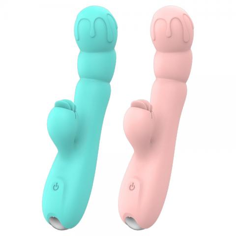
- Vibrators
- Penis Pumps
- Masturbation Cup
- Love Egg
- Contact Us
- [email protected]
- Room 1003, Chevalier House, 188 Chatham Road South, Tsim Sha Tsui, Kowloon, Hong Kong
CopyRight © 2026 wlovew.com. All Rights Reserved.


-
Follow Us On WhatsApp
My Cart (0)
Follow Us On WhatsApp
10$
 Want $10 OFF
Your First Order?
Sign up for the latest product information,offers,and more.
By providing your email address you are consenting to the terms of this privacy policy. select brand exclusions apply.
