Login
 USD $
USD $
 AUD AUD
 CAD CAD
 GBP £
GBP £ EUR €
EUR €
All Categories
(0) My Cart (0)
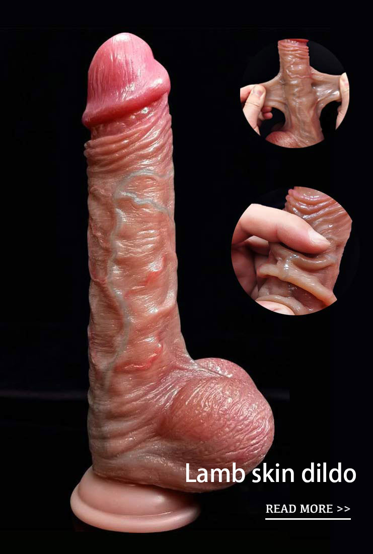
Lambskin Dildo
New Arrival
Mushroom Dildo
Cheap
Huge Dildo
Penis Pumps
Masturbation Cup
Chastity Cage
Pocket Pussy
pleated filter cartridge factory
high flow filter cartridge
large flow filter cartridge
membrane pleated filter cartridge
capsule filter suppliers
capsule filter 0.2 micron
capsule filter price
capsule filter
water filter cartridge


What are the causes of neurosyphilis?
Neurosyphilis (neurosyphilis) is a disease caused by Treponema pallidum (Treponema pallidum) infecting the nervous system, which is divided into congenital and acquired syphilis. So, what are the causes of neurosyphilis?
(1) the cause of the disease
Congenital syphilis is caused by the transmission of syphilis from the mother to the fetus through the placenta, and acquired syphilis patients are mainly infected to each other through sexual behavior.
(2) Pathogenesis
After Treponema pallidum enters the blood, it takes 1 to 3 months to enter the cerebrospinal fluid and invade the central nervous system.
one. Asymptomatic neurosyphilis (asymptomatic neurosyphilis)
The pathological changes of the brain are unknown, because it is difficult for the patients with this disease to obtain autopsy, but it is speculated that most of them mainly involve the meninges, and a few can involve the brain main substance and blood vessels at the same time.
two. Meningeal neurosyphilis (meningeal neurosyphilis)
Pathological changes: although the disease belongs to meningitis, there is often mild cortical damage. Diffuse pial inflammatory reaction, thickening or turbidity can be seen with the naked eye. Miliary gelatinous swelling (gum-ma) can sometimes be seen in the thickened meninges or on the seriously infected meninges, similar to miliary tuberculosis, but the two can be distinguished by microscopic examination.
Microscopic examination showed that the fibrous tissue of the meninges was mainly lymphocyte infiltration, and a small amount of plasma cells vacuum pumps could also be seen. In addition, there could be lymphocyte infiltration around the meningeal vessels, limited inflammatory changes in the convex surface of the brain, and lymphatic and plasma cell infiltration around the Virchow-Robin space.
Meningitis at the base of the brain often damages the brain nerve, showing interstitial inflammatory damage such as eye movement wand vibrator , trochlea and facial nerve. The accumulation of exudates in the base of the brain can block the circulation of cerebrospinal fluid. Even blocking the median foramen or accessory foramen of the fourth ventricle leads to pathological changes of hydrocephalus. The ependymal layer of the ventricular wall is sand-grained or granular, which is due to the proliferation of subependymal astrocytes. If gum-like swelling can be seen in the thickened meninges, it can be seen microscopically that contains fibroblasts, multinucleated giant cells, plasma cells, necrotic tissue in the center, and reticular protein (reticulin) from reticular tissue, which can be distinguished from tuberculosis nodules. Because there is no reticulin in the caseous necrotic tissue of tuberculosis nodules, if the diameter of gum-like swelling is up to a few centimeters, it can oppress adjacent nerve tissue. According to its location and size, it causes different focal symptoms in clinic. in the damaged leptomeninges, sometimes fibrous stellate cells in the cerebral cortex proliferate and extend into the subarachnoid space, meninges and blood vessels of the brain. Endoangitis and extravascular inflammation are common squirting dildo , sometimes leading to encephalomalacia.
Syphilitic meningeal damage, if limited to the spinal cord, is called syphilitic spinal spider reticulitis, and involving the dura mater is called sclerosing meningitis.
three. Pathological changes of vascular neurosyphilis (vascular neurosyphilis):
Vascular neurosyphilis mainly involves the middle and small arteries of the brain and spinal cord, syphilitic endoarteritis and corresponding regional brain and spinal cord tissue malacia.
The common damaged arteries are the recurrent artery of the anterior cerebral artery, which mainly supplies the anterior and inferior part of the head of caudate nucleus, adjacent putamen and anterior limb of internal capsule, middle cerebral artery and its branches, the latter supplying putamen, caudate nucleus, the lower part of the anterior limb of the internal capsule, the dorsal part of the posterior branch of the internal capsule and the globus pallidus, vertebral artery, basilar artery and anterior spinal artery.
The lesion can be limited to an artery or a segment of an artery. The adventitia of the injured artery thickens, lymphocytes and plasma cells infiltrate, the middle layer becomes thinner, the inner muscle layer and elastic fibers are destroyed, but sometimes the elastic fibers remain intact. The thickening of the subintimal fibers narrows the vascular lumen, thrombosis and recanalization in the affected arteries, and some arteries form occlusive endarteritis. Smaller arteries can only have intimal hyperplasia, only narrow the lumen without inflammatory reaction, called Nissl-Alzheimer arteritis, and there may be inflammatory cell infiltration around the nutrient vessels along the wall of large blood vessels. Due to the different vessels of syphilitic arteritis, the location of damaged nerve tissue is different, the size of softening focus is different penis pump , and sometimes there are many small infarctions in the brain.
The above vascular lesions can also be seen in other types of neurosyphilis, such as meningeal vascular syphilis and paralytic dementia. If syphilitic arteritis invades the anterior or posterior spinal artery, it can cause varying degrees of malacia in one or more segments of the spinal cord, which is related to whether the collateral circulation of the spinal cord is good or not. the malacia area shows typical infarct pathological changes, and aneurysms occur in a few cases.
four. Spinal tuberculosis
The pathological changes of the disease are selective degeneration of the nerve fiber bundle, and mainly the posterior root and posterior funiculus of the lumbar, sacral and lower thoracic spinal cord. In general, the spinal pia mater, especially the dorsal spinal cord, becomes thicker and turbid, and the posterior root of the lower spinal cord becomes thinner and flattened. The dorsal spinal cord is significantly involved, and the posterior cord becomes narrower and shrinks. If the optic nerve is involved, the intracranial segment and optic chiasma become thinner. And adhered to the pia mater around it.
Microscopic examination showed that in the early stage of the disease, the spinal pia mater first had inflammatory changes and thickening, mainly lymphocytes and plasma cell infiltration, and mostly confined to the pia mater and cuff of the posterior root, interstitial neuroinflammatory changes in the posterior root, degeneration of nerve fibers in the root, infiltration of inflammatory cells between fibers, demyelination of the posterior funiculus of the spinal cord, especially in thin bundles, myelin disintegration and nerve fiber atrophy In the late stage, the nerve fibers disappeared and astrocytes proliferated, in which part of the connective tissue proliferated, but there was no necrosis and inflammatory reaction. These changes were especially easy to involve the lower thoracic and lumbosacral segments. Sometimes the sympathetic afferent fibers and anterior horn cells were degenerated, and other conduction bundles in the spinal cord were not involved.
In patients with cerebral palsy, the changes of cerebral nerves are the same as those of the posterior root of spinal cord, and the Ⅲ, Ⅳ and Ⅷ nerves are more easily involved. in patients with optic neuritis, the nerve fibers in the optic nerve and optic chiasma are demyelinated, and the inflammatory infiltration is mainly in the periphery. The arachnoid and optic nerve adhere closely and show the changes of optic nerve chiasmatic arachnoiditis.
A very small number of cases mainly involve the cervical spinal cord and its posterior root, which is called cervical spinal cord tuberculosis (tabes dorsalis). The pathogenesis of this disease selectively involving the posterior root and posterior funiculus of the spinal cord has not been elucidated, and it is generally considered to be a toxic degenerative lesion accompanied with reactive inflammatory changes. as for the relationship between this disease and immunology, it is still under study.
five. Paralytic dementia
The pathological changes are mainly the cerebral cortex, which shows that the meninges become turbid and thickened, and can be adhered to the lower cerebral cortex and its external skull, the brain volume shrinks, the cerebral gyrus narrows, the cerebral sulcus widens, and there is more fluid in the sulcus, which is most significant in the prefrontal lobe, followed by the temporal lobe, the cerebral section may be enlarged on both sides of the ventricle, and the ependymal wall of the ventricular system often shows granular reaction. It is caused by granular ependymtis.
Microscopic examination: especially in the frontal lobe and temporal lobe, there was inflammatory cell infiltration around the blood vessels along the Virchow-Robin space and around the meningeal vessels, mainly lymphocytes, plasma cells and phagocytes. The degree of inflammatory changes varied with the stage of the disease and the degree of infection. In the blood vessels in the cortex, there were lymphocytes and plasma cell infiltration in the adventitia, intimal fibroblasts proliferation, and sometimes Treponema pallidum could be found in the vessels. Diffuse degeneration and loss of nerve cells in the cerebral cortex, and reactive astrocytes, especially the proliferation of microglia, glial nodules can be formed in the proliferation of astrocytes, and iron deposition is common around the small blood vessels in the brain.
In the treated cases, there were some effects on the microscopic findings, the inflammatory reaction decreased, even no inflammatory changes were found, and there was no meningeal hyperplasia, only loss of nerve cells and reactive glial hyperplasia in the cerebral cortex.
six. Congenital neurosyphilis
Multiple stillbirth, early pregnancy infection can lead to hydrocephalus and brain development malformation and difficult to survive, early postnatal meningeal vascular infection, adolescent chronic development can show paralytic dementia and spinal cord tuberculosis lesions, its pathological changes are basically the same as adults.
Realistic dildo | best vibrator | pvc dildo | fat pocket pussy | Mushroom Head Dildo | Penis Pumps


- Suction Jelly Dildo
- $6.02

Read More huge dildo
Subscribe for Join Us!
Subcribe to get $10 OFF for order.
- Information
- About us
- Contact us
- Customer Service
- Privacy Policy
- Return Policy
- Shopping Guide
- Payment Methods
- Products
- Dildos
- Vibrators
- Penis Pumps
- Masturbation Cup
- Love Egg
- Contact Us
- [email protected]
- Room 1003, Chevalier House, 188 Chatham Road South, Tsim Sha Tsui, Kowloon, Hong Kong
CopyRight © 2026 wlovew.com. All Rights Reserved.


-
Follow Us On WhatsApp
My Cart (0)
Follow Us On WhatsApp
10$
 Want $10 OFF
Your First Order?
Sign up for the latest product information,offers,and more.
By providing your email address you are consenting to the terms of this privacy policy. select brand exclusions apply.
